Long before the first IVF baby, scientists learned the art in animals: rabbits, hamsters, mice, cattle and sheep. This room tells the story of those discoveries, and of the people and animals behind them.
I
Before Louise: the animal pioneers
The birth of Louise Brown in 1978 was the end of a much longer journey. The fluid her embryo grew in was first perfected for hamster eggs. The dish it grew in was designed for mice. The secret of how sperm become ready to fertilise was discovered in rabbits and rats in 1951. And the very first embryo transfer, moving an embryo from one mother to another, was carried out with rabbits in 1890.
This room tells that story, from Walter Heape's rabbits in 1890 to Frostie, the first calf born from a frozen embryo. There are seventeen discoveries here, and each one links forward to the human work it made possible, so you can follow a single idea from the animal house to the clinic. The story continues in The Founding Papers.
A few famous stories turned out a little differently when we went back to the original papers. Where that happens, we tell you, with the evidence.
And we remember the animals. More than two and a half thousand mice took part in the first embryo-freezing experiments alone [AW·14]. The techniques that now help families around the world were learned first in them.
II
The catalogue
Seventeen objects, 1890 to 1973. Each carries its citation, a statement of what it reports, a curatorial note, a forward link into The Founding Papers where there is a direct line, and the provenance of our own record of it.
AW·011890
Preliminary Note on the Transplantation and Growth of Mammalian Ova within a Uterine Foster-Mother
Walter Heape · surgery by Samuel Buckley
Proceedings of the Royal Society of London, vol. 48, pp. 457–458. Received 12 November 1890. DOI 10.1098/rspl.1890.0053.
What they foundTwo four-cell ova taken from an Angora doe and transferred into the fallopian tube of a Belgian hare doe, which then bore six young — four resembling herself and her mate, and two undoubted Angoras.
Almost every IVF baby born today arrives by the step Walter Heape first showed in 1890. Moving a living embryo from one mother to another, and watching it grow into a healthy young animal, began here, with two tiny Angora embryos and a Belgian hare doe.
The charm of the story is that Heape was not trying to invent anything of the kind. He wanted to know whether a foster-mother could change the nature of the young she carried, one of the great questions about heredity in his day. The answer, he wrote, was no: “So far as this single case goes, the evidence is negative.” Embryo transfer was the happy by-product. He placed the embryos in the fallopian tube, and two Angoras in, two Angoras born, was his proof.
The delicate surgery was done by Samuel Buckley, thanked in the paper's last paragraph and too often forgotten since. You can meet him in section VI.
Leads toThe Founding Papers · FP·04 — Steptoe & Edwards 1978 — a birth whose last step is the one Heape invented
About the sources
DIRECT — full OCR text of Proc. R. Soc. Lond. vol. 48 read from the Internet Archive scan, cross-checked against Crossref metadata and the Royal Society's deposited abstract. Note that Crossref stamps the issue 1891-12-31; that is a metadata artefact and the paper is 1890.
AW·021934
Can Mammalian Eggs Undergo Normal Development in Vitro?
Gregory Pincus · E. V. Enzmann
Proceedings of the National Academy of Sciences USA 1934;20(2):121–122. DOI 10.1073/pnas.20.2.121. Followed by: Pincus G & Enzmann EV, “The Comparative Behavior of Mammalian Eggs in Vivo and in Vitro. I. The Activation of Ovarian Eggs,” Journal of Experimental Medicine 1935;62(5):665–675, DOI 10.1084/jem.62.5.665.
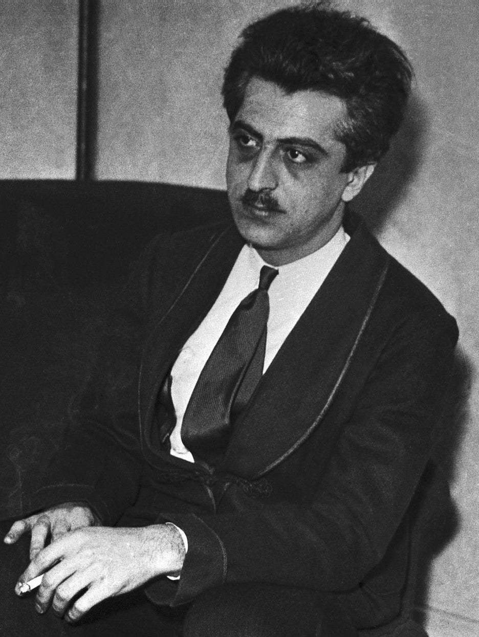
Gregory Pincus in a press photograph of March 1936, issued at the time of his claim of rabbit fertilisation in vitro.ACME, 1936. Public domain in the United States, via Wikimedia Commons.
What they foundThe 1934 paper reports nine live young, in two separate experiments. Ten rabbit ova, recovered from a doe mated to a vasectomised buck, were exposed to sperm for twenty minutes and transferred to a pseudopregnant doe, which produced seven dark grey young thirty-three days later. Separately, five already-fertilised ova were cultured for twenty hours and transferred, giving two young. Pincus and Enzmann claimed “the first certain demonstration that mammalian eggs can be fertilized in vitro”. The 1935 follow-up, which inseminated ovarian eggs in vitro, reports cleavage in a few and polyspermy in the rest — and no births at all.
Gregory Pincus was one of the boldest scientists of his century, and in 1934 he announced that he had fertilised rabbit eggs in a dish. Nine young rabbits were born from his experiments, and the news made him famous.
Looking back, the team could not be sure where fertilisation happened. Sperm carried across with the eggs may have done the work inside the mother rabbit, and twenty minutes with the eggs was far too short for sperm that, as Chang and Austin would discover in 1951 [AW·05], need hours to become ready. That is why the field dates certain mammalian IVF to Chang in 1959.
Pincus's lasting gift lies elsewhere in the same work, and it is a beautiful one: an egg freed from its follicle begins to mature all by itself. “The mere process of explanting ova results in the initiation of maturation.” Every in-vitro maturation since rests on that observation, and it is the very finding Robert Edwards picked up at Mill Hill after 1958 and built his life's work on. Pincus's real contribution to IVF is the egg.
Both papers now DIRECT. The 1935 paper was read via Europe PMC (PMC2133299) when this room opened; the 1934 PNAS note was unobtainable then and this label carried a REGISTRY ONLY flag saying so. It was read in full in August 2026 (PMC1076355), and reading it changed the entry — which is the argument for flagging gaps rather than filling them. One citation correction: the title The comparative behavior of mammalian eggs in vivo and in vitro belongs to the 1935 paper, not to the 1934 one, and the two are routinely welded together in the literature.
AW·031944
In Vitro Fertilization and Cleavage of Human Ovarian Eggs
John Rock · Miriam Friedman Menkin
Science, 4 August 1944; 100(2588):105–107. DOI 10.1126/science.100.2588.105. Full report published four years later with the authors reversed: Menkin MF & Rock J, American Journal of Obstetrics and Gynecology 1948;55(3):440–452, DOI 10.1016/S0002-9378(15)32963-X.
What they foundOf nearly 800 human follicular eggs studied, 138 were exposed to spermatozoa; cleavage to the two- and three-cell stage was observed in three, and interpreted as fertilization.
In wartime Boston, more than thirty years before Louise Brown, John Rock and Miriam Menkin tried the human experiment. Menkin worked patiently through nearly 800 eggs, and in three of them she saw what looked like the first divisions of life.
They were simply too early. In 1944 nobody knew that sperm must be prepared before they can fertilise an egg [AW·05], and the animal groundwork that would make human IVF reliable had not yet been laid. Scientists still debate what Menkin saw, and the result was never repeated.
Their courage still matters. Rock and Menkin showed the world that the question could be asked of human eggs, and Menkin's patient bench work earns her a place among the pioneers.
Leads toThe Founding Papers · FP·04 — Steptoe & Edwards 1978 — what a human claim looks like once the animal work is done
About the sources
REGISTRY for the citation — Crossref and Europe PMC, including the 1948 follow-up and its reversed author order. CONTESTED on the finding. The quoted figures are verbatim from the 1944 paper as reproduced in Thompson 2016 (PMC5991883, open access); Science itself returned 403. The single most valuable source on this question, Biggers' 2012 RBMO review, was unreachable on every route.
AW·041951
Successful Transplantation of a Fertilized Bovine Ovum
E. L. Willett · W. G. Black · L. E. Casida · W. H. Stone · P. J. Buckner
Science, 2 March 1951; 113(2931):247. DOI 10.1126/science.113.2931.247. Followed by Willett EL, Buckner PJ & Larson GL, “Three Successful Transplantations of Fertilized Bovine Eggs,” Journal of Dairy Science 1953;36(5):520–523.
What they foundA single calf produced by transplantation of a fertilized bovine ovum, at Wisconsin. One page.
Heape's rabbit trick, sixty years on, in a cow. One calf, born at Wisconsin in 1950 and reported in March 1951, proved that embryo transfer worked in a large animal.
Embryo transfer is the older half of IVF, and it was the livestock industry, not the clinic, that turned it into a routine procedure. That road led straight to the human embryo transfers of the 1970s.
REGISTRY — Crossref and Europe PMC agree on authors, volume, issue, page and date; the 1953 follow-up title independently corroborates that the 1951 paper reported one calf. Body not read (Science returns 403). Note that Crossref carries three duplicate DOI records for this single one-page paper — a deposit artefact, not three papers.
AW·051951
The co-discovery of capacitation
Min Chueh Chang · Colin Russell Austin
Chang MC, “Fertilizing Capacity of Spermatozoa deposited into the Fallopian Tubes,” Nature 1951;168:697–698, published 20 October 1951, DOI 10.1038/168697b0. · Austin CR, “Observations on the Penetration of the Sperm into the Mammalian Egg,” Australian Journal of Scientific Research, Series B 1951;4(4):581–596, DOI 10.1071/BI9510581. · The coinage: Austin CR, “The ‘Capacitation’ of the Mammalian Sperm,” Nature 1952;170:326, published 23 August 1952, DOI 10.1038/170326a0.
What they foundThat mammalian spermatozoa must spend a period in the female tract, undergoing a physiological change, before they can fertilize an egg. Chang: “a period of time in the female tract is required for the spermatozoa to acquire their fertilizing capacity.”
This is the key that unlocked the whole field. Before 1951, every attempt to fertilise a mammalian egg in a dish began with fresh sperm placed beside an egg, and it never worked. Nobody could see why. Then, on opposite sides of the world, Min Chueh Chang and Colin Russell Austin found the answer: sperm must spend time in the female body, changing, before they can fertilise.
Once the hidden step had a name, it could be met. Within three years Thibault and Dauzier saw a sperm inside a rabbit egg, and within eight Chang had live young [AW·07]. Preparing sperm entirely outside the body came next, in the golden hamster: Yanagimachi and Chang in 1963 [AW·08], then Yanagimachi and Bavister in 1969 [AW·12]. Bavister's medium is the one Edwards used on human sperm that same year.
It is also one of the kindest priority stories in science. Austin coined the word “capacitation” in 1952 and opened his paper by crediting Chang; Chang wrote of its discovery by “Chang and Austin”. The word was born shared.
Leads toThe Founding Papers · FP·01 — Edwards, Bavister & Steptoe 1969 — where human sperm were capacitated in a dish
About the sources
DIRECT for both Nature papers — article pages fetched, titles, volumes, pages, DOIs and publication dates read from the page, opening paragraphs read in full. Austin 1951: REGISTRY + publisher page, CONTESTED on three details — MEDLINE indexes the title as “in the mammalian egg” where the publisher and both Chang's and Yanagimachi's reference lists read “into”; the publisher retrospectively labels the back issue Australian Journal of Biological Sciences, but the as-published 1951 title is Australian Journal of Scientific Research, Series B; and the month is given as November by MEDLINE and December by the publisher. Neither 1951 paper carries an obtainable submission date, so priority by submission is unverified — by publication date Chang was first by four to eight weeks, and that is all the record supports.
AW·061956
Culture of Tubal Mouse Ova — and the discovery that pH was the whole game
Wesley Kingston Whitten · building on John Hammond Jr.
Whitten WK, “Culture of Tubal Mouse Ova,” Nature, 14 January 1956; 177(4498):96, DOI 10.1038/177096a0. Sequel: Whitten WK, “Culture of Tubal Ova,” Nature 1957;179:1081–1082, DOI 10.1038/1791081a0. Predecessor: Hammond J, “Recovery and Culture of Tubal Mouse Ova,” Nature 1949;163(4131):28–29, DOI 10.1038/163028b0.
What they foundEight-cell mouse ova developing consistently to blastocysts in vitro — and the identification of pH drift as the variable that had been blocking it. Ova failed to differentiate above pH 7.7; Krebs–Ringer bicarbonate at pH 7.4 solved it.
Every culture dish warming under carbon dioxide in an IVF laboratory today descends from this one-page letter. Wesley Whitten found that the hidden enemy of the growing mouse embryo was simply pH. Hold the medium at 7.4 and the embryos thrived.
A year later he stripped the recipe down even further, replacing albumin with a pinch of glycine, and concluded that “mouse ova do not require any specific tubal factor at this stage.” There was no secret ingredient in the body to copy. There was only chemistry, and that made growing a human embryo in a dish thinkable.
Leads toThe Founding Papers · FP·06 — Edwards, Steptoe & Purdy 1980 — the culture conditions of the Oldham programme
About the sources
DIRECT — all three Nature article pages fetched and opening paragraphs read; Crossref and Europe PMC cross-checks. These are one-page letters whose opening paragraphs carry the substance; full texts not read. Europe PMC gives Hammond's page as 28, Crossref as 28–29; use 28–29.
AW·06A1958
Successful Development and Birth of Mice cultivated in vitro as Early Embryos
Anne McLaren · John D. Biggers
Nature 1958;182(4639):877–878, 27 September 1958. DOI 10.1038/182877a0. Royal Veterinary College, London.
What they foundMouse embryos recovered at the eight-cell stage, cultured in Whitten's medium for about twenty-four hours until they reached blastocyst, then transferred to the uteri of foster mothers using the embryo-transfer method McLaren had developed with Donald Michie. The recipients carried to term and normal, genetically marked live young were born — the marker being albinism, so that the cultured embryos' offspring could be told from the recipient's own.
Anne McLaren and John Biggers proved something wonderful in 1958: an embryo can grow in glass and still become a healthy baby. They cultured mouse embryos for a day, placed them in foster mothers, and watched normal young arrive, each carrying a coat-colour marker that proved it came from a cultured embryo.
IVF is a chain of steps: an egg, fertilisation outside the body, growth outside the body, transfer, birth. Heape had shown transfer in 1890 [AW·01], and Chang would show fertilisation in 1959 [AW·07]. This paper, a year before Chang, showed that time in a dish does not cost an embryo its future.
It was exactly what Robert Edwards needed. In the early 1960s he set out to learn about culture media for his eggs and embryos, with “successful mouse embryo culture only recently having been described (McLaren and Biggers, 1958).” Here the embryos were fertilised inside the mother and only grown outside, a detail sometimes lost in retellings. The modest authors themselves credited C. E. Adams with combining culture and transfer first, in rabbits, but it was McLaren and Biggers who brought the babies.
Leads toThe Founding Papers · FP·01 — Edwards, Bavister & Steptoe 1969 — human eggs matured and fertilised in culture
About the sources
PARTIAL. Citation, title, date, DOI, affiliation and the paper's own opening read DIRECT from Nature; the full text is paywalled and was not read. The experimental particulars — eight-cell stage, Whitten's medium, roughly twenty-four hours, albino markers — are corroborated from three independent sources including Papaioannou VE, Int J Dev Biol 2001;45(3):483–486, whose Figure 1 caption reads: “The first adult mice produced by embryo transfer in which part of the preimplantation stages of development occurred in vitro.” Note that PubMed carries a corrupted form of the title — use the Nature form.
AW·071959
Fertilization of Rabbit Ova in vitro
Min Chueh Chang
Nature, 8 August 1959; 184(4684):466–467. DOI 10.1038/184466a0. Immediate prior art, cited by Chang himself: Dauzier L, Thibault C & Wintenberger S, Comptes Rendus de l'Académie des Sciences 1954;238:844–845 — cytological evidence of rabbit ova fertilized in vitro using capacitated sperm.
What they foundA repeatable procedure for fertilizing rabbit ova in vitro, and the normal development in vivo of ova so fertilized after transfer to recipient rabbits.
The moment mammalian IVF became real. Min Chueh Chang knew that seeing a sperm in an egg under the microscope was not enough, so he set himself the highest test: transfer the fertilised eggs and wait for living young. Black rabbit eggs, fertilised in a dish and carried by a white doe, gave a black litter. There was no arguing with that.
He was generous about those who came close before him, crediting Thibault and Dauzier's 1954 work. And there is a lovely twist in the tale: Chang had been brought to the Worcester Foundation in 1945 by Gregory Pincus, whose bold 1934 rabbit claim [AW·02] this paper finally made good on.
Leads toThe Founding Papers · FP·03 — Edwards, Steptoe & Purdy 1970 — fertilization and cleavage, in the human
About the sources
DIRECT for citation, date, affiliations, opening paragraph and full reference list — Nature article page read. PARTIAL on the outcome numbers: the body is paywalled and we could not establish how many ova were fertilized or how many young were born. Every route was blocked. We print no number. The coat-colour genetic control described in tertiary sources is not confirmed. Ignore Europe PMC's “184(Suppl 7)” — a MEDLINE artefact; the volume is 184. One qualification the common accounts elide: Chang's sperm were capacitated in vivo, in the female tract. Capacitation in the dish came later.
AW·081963
Fertilization of Hamster Eggs in vitro
Ryuzo Yanagimachi · Min Chueh Chang
Nature, October 1963; 200(4903):281–282, DOI 10.1038/200281b0. Full paper: “In vitro fertilization of golden hamster ova,” Journal of Experimental Zoology 1964;156(3):361–375, DOI 10.1002/jez.1401560312.
What they foundIn-vitro fertilization of golden hamster ova.
Meet the golden hamster, the little animal that carried human IVF over the line. Ryuzo Yanagimachi and Min Chueh Chang fertilised hamster eggs in a dish in 1963, and the hamster became the workbench of fertilisation research.
It was the hamster system that Barry Bavister perfected [AW·12], and a hamster medium that made the first human fertilisation possible in 1969. Hamster embryos themselves grew poorly beyond two cells, and the mouse work filled that gap. For years afterwards the hamster egg test was also used in clinics to check human sperm.
Leads toThe Founding Papers · FP·01 — Edwards, Bavister & Steptoe 1969 — the species that supplied its medium
About the sources
REGISTRY — both DOIs, volumes, pages and PMIDs confirmed in Crossref and Europe PMC. No abstracts are indexed for either paper and neither body was read — PARTIAL on content. Yanagimachi's own 2022 retrospective was retrieved but does not narrate this episode.
AW·091963
A Method for in vitro cultivation of mouse ova from two-cell to blastocyst
Ralph Lawrence Brinster
Experimental Cell Research, October 1963; 32(1):205–208. DOI 10.1016/0014-4827(63)90093-4. With the systematic series: “Studies on the development of mouse embryos in vitro. I–III,” J Exp Zool 1965;158(1):49–57, 59–68, 69–77; and “IV. Interaction of energy sources,” J Reprod Fertil 1965;10(2):227–240.
What they foundA micro-drop-under-oil method for culturing two-cell mouse ova to blastocyst, followed by a four-part factorial dissection of the variables that govern it: osmolarity, hydrogen ion concentration, energy source and fixed-nitrogen source.
Look into any IVF culture dish today and you are looking at Ralph Brinster's idea: tiny drops of medium under a protective layer of oil. His careful series of papers worked out the saltiness, the pH and the energy sources an embryo needs, and those values still shape every clinical culture medium.
This was a way of growing embryos, not of fertilising them, and it is one of the quiet foundations every IVF laboratory stands on.
REGISTRY — all five DOIs and paginations confirmed in Crossref and Europe PMC. Article bodies not read, so the description of the factorial design rests on titles and registry abstracts — PARTIAL on content.
AW·101968
Complete development in vitro of the pre-implantation stages of the mouse in a simple chemically defined medium
Wesley Kingston Whitten · John Dennis Biggers
Journal of Reproduction and Fertility 1968;17(2):399–401. DOI 10.1530/jrf.0.0170399. The named formulations that followed: Whitten's medium (Advances in the Biosciences 6, 1971, pp. 129–141) and BWW — Biggers, Whitten & Whittingham, “The culture of mouse embryos in vitro,” in Daniel JC Jr (ed.), Methods in Mammalian Embryology, Freeman, 1971, pp. 86–116.
What they foundMouse embryos carried through the entire preimplantation period in vitro in a simple chemically defined medium — no serum, no egg white, no biological fluid of unknown composition.
The end of a long road that began with egg white. In 1968 Wesley Whitten and John Biggers carried mouse embryos through their whole first week in a simple, fully defined medium, with nothing mysterious added at all.
David Whittingham used exactly this medium for mouse IVF the same year [AW·11]. From this group came BWW, the sperm medium of the early IVF laboratories, and Whitten's medium, ancestor of the mouse embryo test that still checks every batch of human IVF culture medium. The mouse became the field's quality control.
Leads toThe Founding Papers · FP·01 — Edwards, Bavister & Steptoe 1969 — the medium Whittingham cited, one year on
About the sources
Whitten & Biggers 1968 and Whitten 1971: REGISTRY (Crossref, Europe PMC). BWW 1971: SEARCH-VERIFIED — a book chapter with no DOI, verified through the Jackson Laboratory institutional bibliography and corroborated by independent secondary citations of identical pagination. It is the most-cited item in this room with the weakest machine-readable provenance, and the chapter does not call itself “BWW” — that is a community coinage applied later.
AW·111968
Fertilization of Mouse Eggs in vitro
David Gordon Whittingham
Nature 1968;220(5167):592–593. Received 8 August 1968; revised 30 September 1968; published 9 November 1968. DOI 10.1038/220592a0.
What they foundIn the author's words: “the in vitro fertilization of mouse eggs in a simple chemically defined medium, and the subsequent development of the fertilized eggs to 17 day old foetuses.”
In 1968, three years earlier than many books say, David Whittingham fertilised mouse eggs in a simple defined medium and saw them grow into healthy late-stage fetuses. From this moment the mouse became the model animal of assisted reproduction.
The 1971 date often quoted belongs to a follow-up paper by Iwamatsu and Chang. The rat followed soon after, and it was in rats that Miyamoto, Toyoda and Chang worked out how to prepare sperm fully in a dish (1973 to 1974). Human IVF needs exactly that step, and this is where the recipe was written.
DIRECT — Nature article page fetched with received, revised and published dates and abstract text read; Crossref and Europe PMC corroboration. All competing priority claims registry-verified with their own dates. Note the eighteen-month gap between receipt and publication of Miyamoto & Chang — any priority argument in this era that uses publication dates rather than receipt dates will go wrong.
AW·121969
Environmental factors important for in vitro fertilization in the hamster
Barry D. Bavister
Journal of Reproduction and Fertility, April 1969; 18(3):544–545. DOI 10.1530/jrf.0.0180544. The author's own retrospective: Bavister BD, “How animal embryo research led to the first documented human IVF,” Reproductive BioMedicine Online 2002;4(Suppl 1):24–29, DOI 10.1016/s1472-6483(12)60008-x.
What they foundThat hamster fertilization in vitro is steeply pH-dependent — at pH 7.2 or below almost no oocytes were penetrated; between 7.3 and 7.5, forty per cent were fertilized; at 7.6 and above, more than seventy-five per cent. Sperm motility was unaffected. The finding produced a culture medium, Tyrode-B.
Here the animal story walks straight into the human one. Barry Bavister, a young researcher in Cambridge, discovered that hamster fertilisation depends on pH: below 7.2 almost nothing happened, while above 7.6 more than three quarters of the eggs were fertilised. His new medium, Tyrode-B, was the result.
Then it was tried on human eggs. In Bavister's own words: “When this medium was used for human gametes, IVF was observed and documented for the first time. Spermatozoa penetrating through the zona pellucida and sperm components within the ooplasm were detected, and some oocytes exhibited two pronuclei.”
That is why his name sits in the middle of the famous 1969 paper: Edwards, Bavister and Steptoe. He told this story himself, many years later.
Leads toThe Founding Papers · FP·01 — Edwards, Bavister & Steptoe 1969 — the paper this medium made possible
About the sources
The 1969 note: REGISTRY (Crossref, Europe PMC); body not retrieved, and we could not determine whether it is a full short communication or a meeting abstract — it is two pages. The 2002 retrospective: REGISTRY, full abstract retrieved verbatim from Europe PMC. The load-bearing claim rests on a participant's account written thirty-three years after the fact. It is first-person testimony by a named author of the 1969 paper, published in a peer-reviewed journal — strong, but testimony. We could not retrieve the body of the 1969 Nature paper to confirm the medium is named in its methods. If the 1969 note is indeed only an abstract, then the founding dataset behind human IVF's culture medium sat in a Cambridge PhD thesis and two pages of a fertility journal for three decades. That would be a remarkable fact, and we cannot confirm it.
AW·131972
A two-factor hypothesis of freezing injury
Peter Mazur · Stanley P. Leibo · Ernest H. Y. Chu
Experimental Cell Research, April 1972; 71(2):345–355. Subtitle: “Evidence from Chinese hamster tissue-culture cells.” DOI 10.1016/0014-4827(72)90303-5.
What they foundThat freezing injury has two distinct causes which act in opposite directions as cooling rate changes — evidenced in Chinese hamster tissue-culture cells.
Why do some cells survive freezing and others die? In April 1972 Peter Mazur, Stanley Leibo and Ernest Chu gave the answer. Cool a cell too slowly and the salts left behind as water escapes injure it. Cool it too fast and the water freezes inside. Between the two lies a sweet spot.
Every slow-freezing method in embryology is a search for that sweet spot, and vitrification, which came later, is a clever escape from the problem altogether. The physics came six months before the famous mouse embryo paper, by two of the same authors.
REGISTRY — Crossref and Europe PMC agree on the full title including the subtitle, which is usually dropped in citation, plus volume, issue, pages and date. Body not read; the mechanism described above is standard cryobiology rather than a claim read off this paper. PARTIAL on the paper's specific argument. Its evidence is tissue-culture cells, not embryos — the extension to embryos is inference, well-founded and made by the same people, but inference.
AW·141972
Survival of Mouse Embryos Frozen to −196° and −269°C
David Gordon Whittingham · Stanley P. Leibo · Peter Mazur
Science, 27 October 1972; 178(4059):411–414. DOI 10.1126/science.178.4059.411. Preceded by Whittingham DG, “Survival of Mouse Embryos after Freezing and Thawing,” Nature 1971;233(5315):125–126, received 17 May 1971, DOI 10.1038/233125a0.
What they foundFrom the abstract, verbatim: “Depending on the specific rates used, 50 to 70 percent of more than 2500 frozen and thawed early embryos developed into blastocysts in culture after storage at −196°C for up to 8 days. When approximately 1000 of the survivors, including some frozen to −269°C (4°K), were transferred into foster mothers, 65 percent of the recipients became pregnant. More than 40 percent of the embryos in these pregnant mice gave rise to normal, living full-term fetuses or newborn mice.”
The birth of embryo freezing. At Oak Ridge National Laboratory, David Whittingham, Stanley Leibo and Peter Mazur froze more than 2,500 mouse embryos, some to just four degrees above absolute zero, thawed them, and transferred about a thousand to foster mothers. Healthy mice were born.
This is the ancestor of the frozen embryo transfer, now the majority of IVF transfers worldwide. It makes possible single embryo transfer, deferred transfer and fertility preservation: everything that depends on an embryo being able to wait.
Whittingham had already been working on the problem for over a year, publishing in Nature in 1971 that mouse embryos could survive freezing. This paper added the babies.
REGISTRY — Crossref for volume, issue, pages and date; Europe PMC for the PMID and the full abstract, retrieved verbatim, which is the source of every number quoted above. No printed received date obtainable; science.org returns 403. The −79°C detail sometimes given for the 1971 paper is search-verified only and we do not print it.
AW·151972
The effect of cooling rate, warming rate, cryoprotective agent and stage of development on survival of mouse embryos during freezing and thawing
Ian Wilmut
Life Sciences Part 2, November 1972; 11(22):1071–1079. DOI 10.1016/0024-3205(72)90215-9. Companion: Wilmut I, “The low temperature preservation of mammalian embryos,” J Reprod Fertil 1972;31(3):513–514.
What they foundThe effect of four variables — cooling rate, warming rate, choice of cryoprotectant and embryo stage — on mouse embryo survival through freeze and thaw.
The same discovery, made at the same time, in two places. While the Oak Ridge team froze mouse embryos in Tennessee, Ian Wilmut in Cambridge was independently working out the same four things: how fast to cool, how fast to warm, which protective liquid to use and which embryo stage to freeze.
Two laboratories reaching the same answer shows the method was written by nature, not by luck. And yes, this is the same Ian Wilmut later famous for Dolly the sheep. His achievement here stands on its own: a frozen mammalian embryo can become a living animal.
REGISTRY — Crossref and Europe PMC agree on volume, issue, pages and month. No received or submitted date is obtainable: Elsevier's 1972 deposits carry no received-date assertion, ScienceDirect is robots-disallowed, and no repository or archive copy exists. The printed “Received” line, which papers of this era carried, is therefore unread. Until someone puts eyes on a physical copy of Life Sciences 11(22), this room cannot say who submitted first — only who appeared in print first. Note also that Crossref's stored title contains a transcription error, “of survival” for “on survival”; Europe PMC has it right.
AW·161973
Frostie — the first calf from a frozen embryo
Ian Wilmut · Lawrence Ernest Arthur Rowson
“Experiments on the low-temperature preservation of cow embryos,” The Veterinary Record, 30 June 1973; 92(26):686–690, DOI 10.1136/vr.92.26.686. Announced first in Wilmut I & Rowson LEA, “The successful low-temperature preservation of mouse and cow embryos,” J Reprod Fertil 1973;33(2):352–353. Third species: Willadsen SM, Polge C, Rowson LEA & Moor RM, “Deep freezing of sheep embryos,” J Reprod Fertil 1976;46(1):151–154, DOI 10.1530/jrf.0.0460151.
What they foundBlastocysts frozen and stored six days in liquid nitrogen, thawed, cultured overnight and surgically transferred to synchronised recipients — producing a Hereford–Friesian cross bull calf that Wilmut named Frostie.
Meet Frostie, the first calf ever born from a frozen embryo. In 1973 Ian Wilmut and Lawrence Rowson froze a cattle embryo in liquid nitrogen for six days, warmed it, and transferred it. The Hereford-Friesian bull calf that followed was named Frostie, and he turned embryo banking from a laboratory curiosity into a technology.
The sheep work that followed showed that the speed of warming matters as much as the speed of cooling, a lesson human freezing still follows. In a charming touch of the era, the embryos were first tested by incubating them inside rabbit oviducts. And cattle gave IVF one more gift: the first calf conceived by IVF was born on 9 June 1981.
REGISTRY for both 1973 citations and for the 1976 sheep paper, whose full abstract was retrieved verbatim — every sheep number above comes from the source. The calf's breed, six-day storage and overnight culture are DIRECT from the 2023 Reproduction obituary of Wilmut. The name “Frostie” is search-verified from the University of Edinburgh Library's archival material drawing on the Wilmut papers, and appears in retrospective accounts rather than necessarily in the 1973 paper itself. Bodies of the 1973 papers not read. Iritani & Niwa 1977 is the weakest verification in this room — no abstract is indexed anywhere and the body was unreachable, so that entry rests on its title alone, which is why it is a note here rather than an object.
III
The famous firsts, at a glance
A quick guide to the milestones in this room. Tap any object number to jump to its full story.
The famous firsts, and where to find them in this room
Two Angora embryos placed in the fallopian tube of a Belgian hare doe; six young born, two of them Angoras. Heape was actually studying heredity, and found embryo transfer along the way.
Pincus and Enzmann's lasting discovery: an egg released from its follicle begins to mature by itself. Their bold rabbit IVF claim was made good by Chang in 1959.
Frostie, a Hereford-Friesian bull calf, from an embryo stored six days in liquid nitrogen.
IV
Two discoveries made twice
Twice in this story, two teams on opposite sides of the world made the same discovery at almost the same moment. Both times, they met it with generosity.
Capacitation, 1951: Austin and Chang
M. C. Chang's note in Nature appeared on 20 October 1951. Colin Austin's paper from Australia followed a few weeks later, in November or December. Neither paper records when it was submitted, so nobody can say who began first.
What the record does show is friendship. By August 1952 Austin's paper naming the process opened by crediting Chang's rabbit findings alongside his own. Chang returned the courtesy in 1959, writing of the recognition of capacitation “by Chang and Austin.” Two scientists, two hemispheres, one discovery, each crediting the other. The word was born already shared.
Embryo freezing, 1972: Oak Ridge and Cambridge
Science published David Whittingham, Stanley Leibo and Peter Mazur on 27 October 1972. Life Sciences published Ian Wilmut about a month later, in November 1972. Wilmut's submission date has not survived, so who finished first remains an open question.
Both teams were guided by Mazur's two-factor theory of freezing injury, published that April [AW·13], and both arrived at the same four controls, because those are the ones the physics of freezing allows. That two teams found the same answer independently is the best sign that the answer was right.
V
Gregory Pincus and the rabbits
In 1934 Gregory Pincus announced that rabbit eggs had been fertilised in a dish. It made headlines, and the story has been repeated ever since. The real story is more interesting.
His 1934 paper with E. V. Enzmann reports nine live young rabbits. Seven came from eggs exposed to sperm for twenty minutes and then transferred to a doe; two came from eggs that were already fertilised before they were cultured.
What the experiment could not rule out was that sperm carried across with the eggs fertilised them inside the doe, rather than in the dish. Twenty minutes was also far too short: as Chang and Austin would discover in 1951 [AW·05], sperm need hours inside the female before they can fertilise at all. The follow-up paper of 1935 went no further than eggs that had begun to divide, many of them entered by more than one sperm, and reported no births.
Pincus's lasting discovery in this work was a different one, and it still matters in every IVF laboratory today: an egg taken from its follicle begins to ripen by itself.
His colleagues were cautious from the start. Austin called the evidence for mammalian fertilisation in vitro “inconclusive” in 1951, and Chang, whom Pincus himself had brought to the Worcester Foundation, wrote in 1957: “up till now we still do not have a repeatable procedure to fertilize mammalian eggs in vitro.” It was Chang who finally did it, in 1959, with live young [AW·07].
Pincus went on to help develop the oral contraceptive, and his fame kept the 1934 story alive. His real gift to IVF is quieter, and it lasted.
VI
The hands that solved it
Behind many of these discoveries were people whose hands did the work, and whose names were too often left out. This museum remembers them.
In 1890 the surgery that made the first mammalian embryo transfer possible was performed by Samuel Buckley, of Manchester. He is named once, in the closing paragraph of Heape's paper [AW·01], and almost never since.
In 1944 Miriam Friedman Menkin ran the bench work of the Free Hospital for Women's long search, working through nearly eight hundred eggs, one hundred and thirty-eight of them exposed to sperm, over several years. On 3 February 1944 she changed the method: one wash instead of three, a more concentrated sample, and a full hour of exposure instead of half. Whatever the result, the change that produced it was hers.
The credit fell like this. The short, famous paper in Science, August 1944: Rock and Menkin. The full thirteen-page report four years later: Menkin and Rock. TIME called her “his assistant”. She had been publishing in the Journal of Experimental Medicine since 1930.
And in 1978 Jean Purdy, who ran the laboratory at Oldham and was a named author on at least eleven research papers with Edwards and Steptoe, was left off the single page that announced Louise Brown's birth. Her story is told on her own page.
Remembering the hands
Again and again, the credit went to the person who asked the question, and not to the person at the bench who answered it. Three times across eighty-eight years, in three countries. This museum names them.
VII
Sources and notes
Every object in this room was checked in August 2026 against the original publication, a bibliographic registry, a digitised archive scan or a library record. Each object's label says how far that check went.
Notes on the documents we could not reach
The numbers in Chang 1959: how many eggs and how many young. The full text is behind a paywall on every route, so we print no number.
Wilmut's 1972 submission date, which is why section IV leaves the order open.
Whether the 1969 Nature paper names Bavister's medium in its methods. This rests on Bavister's own later account.
Iritani and Niwa 1977 on cattle IVF: no abstract is indexed and the text was unreachable, so it appears as a note under AW·16.
The parthenogenesis explanation for Rock and Menkin: often repeated, but we could not verify it, so we do not assert it.
Read as citations only: Yanagimachi and Chang 1963 and 1964; the Brinster 1965 series; both Wilmut and Rowson 1973 papers; Willett 1951; Heape's 1897 follow-up; the BWW chapter, which is not digitised.
Small citation errors in the databases, recorded so they stop spreading: Crossref holds three duplicate DOIs for Willett 1951, reads “of survival” for Wilmut 1972 where the paper reads “on”, and dates Heape 1890 as 1891; MEDLINE reads “in the mammalian egg” for Austin 1951 where the publisher reads “into”; Europe PMC records Chang 1959 as “184(Suppl 7)”.
Search our store
Back to top
Spend $250 to get free shippingSpend $250 more to get free shippingCongratulations! You get free shipping.